Back Muscles Diagram - Creatine is now proving to be one of the most potent muscle growth accelerators giving excellent muscle mass increase and phenomenal strength increases order yours today.
Back Muscles Diagram - Creatine is now proving to be one of the most potent muscle growth accelerators giving excellent muscle mass increase and phenomenal strength increases order yours today.. People with back pain people who experience headaches printing for use during doctor visits to communicate information about your symptoms quickly tracking your progress over time related tools: When back development is the goal, stick to one of these variations. Nerves in your lower back. Some of the links in the post above are affiliate links.. This muscle is a major generator of lower back and hip pain, as well as being responsible for complaints of a burning sensation along the posterior superior iliac spine (psis) and sacroiliac joint.
We think this is the most useful anatomy picture that you need. This human anatomy module is composed of diagrams, illustrations and 3d views of the back, cervical, thoracic and lumbar spinal areas as well as the various vertebrae. Muscles labeled front and back 12 photos of the muscles labeled front and back muscle diagram labeled front and back, muscle system labelling (front and back), muscular system labeled front and back, human muscles, muscle diagram labeled front and back, muscle system labelling (front and back), muscular system labeled front and back. Muscle anatomy crossword answer 12 photos of the muscle anatomy crossword answer muscle anatomy crossword answer key biology corner, muscle anatomy crossword answers biology corner, muscle anatomy crossword answers key, muscle anatomy crossword puzzle answers biology corner, muscle anatomy. These muscles include the large paired muscles in the lower back, called erector spinae, which help hold up the spine, and gluteal muscles.
Back Anatomy All About The Back Muscles from www.kingofthegym.com Stand behind the barbell with your feet shoulder. This diagram depicts muscles in back diagram.human anatomy diagrams show internal organs, cells, systems, conditions, symptoms and sickness information and/or tips for healthy living. We hope this picture anatomy of back muscles diagram can help you study and research. Daniel nelson on january 1, 2019 2 comments 🔥! What is the origin and insertion of the rhomboid minor and major muscle? Extrinsic and intrinsic.the back functions are many, such as to house and protect the spinal cord, hold the body and head upright, and adjust the movements of the upper and lower limbs. Related posts of muscles of the lower back and buttocks diagram muscle anatomy crossword answer. Major muscles back muscles shoulder muscles supraspinatus muscle back workout routine sternocleidomastoid muscle muscle diagram body diagram latissimus dorsi.
Nerves in your lower back. This muscle is a major generator of lower back and hip pain, as well as being responsible for complaints of a burning sensation along the posterior superior iliac spine (psis) and sacroiliac joint. These muscles include the large paired muscles in the lower back, called erector spinae, which help hold up the spine, and gluteal muscles. Major muscles back muscles shoulder muscles supraspinatus muscle back workout routine sternocleidomastoid muscle muscle diagram body diagram latissimus dorsi. Back to tracking tools main page. It is particularly interesting for physiotherapists. This is a diagram of the larger and more surface muscles of the low back. The back muscles enable you to stand up straight; Learn vocabulary, terms, and more with flashcards, games, and other study tools. The extrinsic (superficial) back muscles, which lie most superficially on the back. This diagram depicts muscles in back diagram.human anatomy diagrams show internal organs, cells, systems, conditions, symptoms and sickness information and/or tips for healthy living. See related links to what you are looking for. Support and protect your spine;
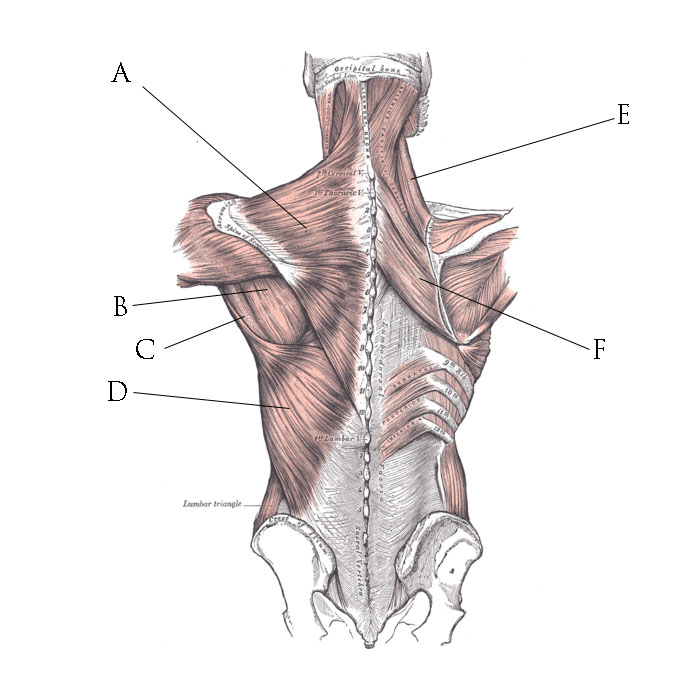
The deltoid, teres major, teres minor, infraspinatus, supraspinatus (not shown) and subscapularis muscles (not shown) all extend from the scapula to the humerus and act on the shoulder joint. The back has a total of 40 muscles. Muscle anatomy crossword answer 12 photos of the muscle anatomy crossword answer muscle anatomy crossword answer key biology corner, muscle anatomy crossword answers biology corner, muscle anatomy crossword answers key, muscle anatomy crossword puzzle answers biology corner, muscle anatomy. For more anatomy content please follow us and visit our website: It is opposite from the chest, and the vertebral column runs down the back.
Back Muscles Diagram Photos And Premium High Res Pictures Getty Images from media.gettyimages.com The muscles of the back can be arranged into 3 categories based on their location: Muscles labeled front and back 12 photos of the muscles labeled front and back muscle diagram labeled front and back, muscle system labelling (front and back), muscular system labeled front and back, human muscles, muscle diagram labeled front and back, muscle system labelling (front and back), muscular system labeled front and back. When back development is the goal, stick to one of these variations. Symptoms of muscle pain include: Some of the links in the post above are affiliate links.. Rotator cuff muscles and tendons. Others, like sumo deadlifts, have been shown in emg studies—and in the trenches—to focus more on other muscle groups than the back. Related posts of muscles of the lower back and buttocks diagram muscle anatomy crossword answer.
Below you'll see diagrams along with the names of the back muscles that may be the cause of your pain.
Five pairs of lumbar spinal nerves labeled l1 to l5 branch off your spinal cord and exit through small holes between the vertebrae. Chronic back pain map this tool recommended for: This diagram depicts muscles in back diagram.human anatomy diagrams show internal organs, cells, systems, conditions, symptoms and sickness information and/or tips for healthy living. Daniel nelson on january 1, 2019 2 comments 🔥! By the way, have you heard about the myth of. Extrinsic and intrinsic.the back functions are many, such as to house and protect the spinal cord, hold the body and head upright, and adjust the movements of the upper and lower limbs. The deltoid, teres major, teres minor, infraspinatus, supraspinatus (not shown) and subscapularis muscles (not shown) all extend from the scapula to the humerus and act on the shoulder joint. Pain log more pain mapping tools Anatomical diagrams of the spine and back. Stand behind the barbell with your feet shoulder. The muscles of the back can be arranged into 3 categories based on their location: This human anatomy module is composed of diagrams, illustrations and 3d views of the back, cervical, thoracic and lumbar spinal areas as well as the various vertebrae. This muscle is a major generator of lower back and hip pain, as well as being responsible for complaints of a burning sensation along the posterior superior iliac spine (psis) and sacroiliac joint.
The most common type of back pain is muscle pain—also called muscle strain or soft tissue strain. This muscle is a major generator of lower back and hip pain, as well as being responsible for complaints of a burning sensation along the posterior superior iliac spine (psis) and sacroiliac joint. How many muscles are in the back? Most of the time, back muscle pain is diagnosed then treated with little more than a prescription of rest, painkillers and muscle relaxants. Learn vocabulary, terms, and more with flashcards, games, and other study tools.
File Back Muscles Jpg Wikimedia Commons from upload.wikimedia.org Most of the time, back muscle pain is diagnosed then treated with little more than a prescription of rest, painkillers and muscle relaxants. For more anatomy content please follow us and visit our website: The pelvis at the bottom of the back and the shoulders at the top of the back give the back. The human back extends from the buttocks to the posterior portion of the neck and shoulders. Below you'll see diagrams along with the names of the back muscles that may be the cause of your pain. What is the origin and insertion of the rhomboid minor and major muscle? Five pairs of lumbar spinal nerves labeled l1 to l5 branch off your spinal cord and exit through small holes between the vertebrae. To learn more about the anatomy of the spine, watch this video.
When back development is the goal, stick to one of these variations.
Back to tracking tools main page. Anatomynote.com found anatomy of back muscles diagram from plenty of anatomical pictures on the internet. This human anatomy module is composed of diagrams, illustrations and 3d views of the back, cervical, thoracic and lumbar spinal areas as well as the various vertebrae. Five pairs of lumbar spinal nerves labeled l1 to l5 branch off your spinal cord and exit through small holes between the vertebrae. Back muscles, back muscle diagram. What is the origin and insertion of the rhomboid minor and major muscle? Some of the links in the post above are affiliate links.. This is a diagram of the larger and more surface muscles of the low back. For more anatomy content please follow us and visit our website: Rotator cuff muscles and tendons. Muscle spasms (contraction or stiffening of the back muscles) muscles that feel tight; Both the deltoid and the trapezius are firmly attached to the spine of the scapula. These muscles include the large paired muscles in the lower back, called erector spinae, which help hold up the spine, and gluteal muscles.


0 Komentar